The Truth About…Cataracts
At least a couple times a week someone brings in an older dog and tells me the pet is getting cataracts. Fortunately, the changes they’ve noticed usually aren’t true cataracts and don’t significantly trouble their vision. True cataracts do happen in pets, though, and not uncommonly either. When cataracts are large or progressive they can lead to blindness or even painful glaucoma, so it’s important they are recognized and closely monitored by your veterinarian.

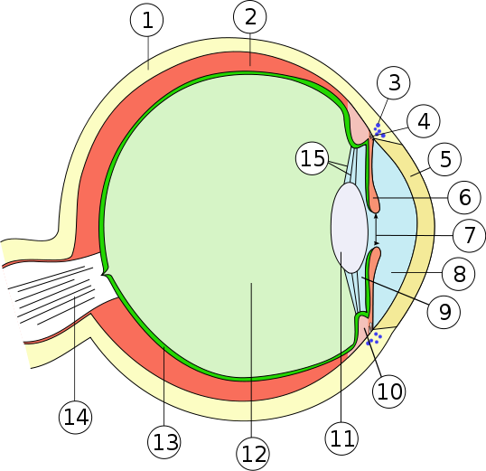
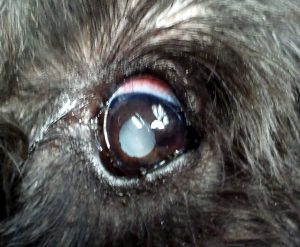
Cataracts are imperfections in the lens – the clear disk which sits in the pupil and focuses light on the back of the eye, which in turn translates that light into an image*. So, the important thing about the lens is that it is clear and flexible (little muscles attach that deform it to focus the light so you can see near and far). There is a very intricate, highly organized structure to the proteins in the lens that allows this to happen. A cataract, then, occurs when this structure breaks down, turning part or all of the lens opaque. Light can no longer pass through that area, interfering with vision to a degree dependent on the size of the cataract. A small cataract may have no significant impact on vision, but complete (or mature) cataract essentially means that eye is blind.

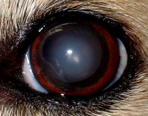
is central and surrounded by
normally transparent lens.
Before we go any further, let’s clarify whatmost owners get concerned about, a phenomenon called nuclear sclerosis – which thankfully doesn’t have a noticeable effect on vision and isn’t a true cataract. As dogs age, new lens fibers slowly develop from the outside edge and move inward, slowly compressing the center (nucleus) of the lens. The increased density results in a hazy appearance that is often confused with a cataract, but unlike a cataract light still passes through fairly normally. Indeed, one way we differentiate these from true cataracts is by shining a bright light into the eye; with nuclear sclerosis you’ll see the iridescent reflection from the back of the eye, but with a cataract you won’t. Since light still passes through, vision is still effective – perhaps it’s a bit blurry, but I’ve yet to see a pet noticeably effected by it.
So, what can cause the delicate, highly-specialized lens structure to break down and lose transparency (i.e., form a cataract)? Genetics can certainly play a role, and there’s a laundry list of breeds predisposed to cataract formation. Sometimes these are present at birth and sometimes they develop with age.

opaque lens.
Many other problems and diseases may cause cataracts as a secondary consequence – basically, anything that disrupts the environment within the eye can lead to cataract formation. Inflammation inside the eye (or uveitis) is a common culprit, and due to some quirks of structure the eye is a common place for inflammation to develop even when problems start elsewhere. This happens with many bacterial infections and autoimmune diseases, and it’s another reason a complete physical exam by a veterinarian is so important in every animal -sometimes a good eye exam leads to discovering another problem, or vice-versa. Trauma to the eye, or (yikes) to the lens itself can certainly result in cataracts as well.
Vision loss is not the only potentially serious consequence of cataracts, however. When progressive, the lens may break down to the point that it leaks proteins into the rest of the eye, causing serious inflammation and even glaucoma – an extremely painful condition where pressure builds up inside the eye. Fortunately many cataracts are not progressive, but for this reason they should all be closely monitored by your veterinarian. Diabetic dogs merit special mention, as most of them will develop rapidly progressive cataracts due to differences in the way their lenses metabolize sugar. Considering this, all owners of newly diagnosed diabetic dogs get educated on how and why to monitor for cataracts. Likewise, any dog that suddenly develops progressive cataracts should be screened for diabetes.
While there is no proven medical treatment to slow or reverse cataracts, the same surgical procedure used in humans may be performed in pets. This involves breaking up the material in the lens, suctioning it out, and placing a prosthetic lens inside the remaining capsule. Obviously, it’s a delicate procedure that requires lots of special (not to mention expensive) equipment and training, so it is only performed by veterinary ophthalmologists. Your family veterinarian should be able to direct you to a specialist and give you an idea of the costs in your area. The procedure has a high success rate for returning vision, but not everyone will be able to realistically afford the expense. Not every pet is a candidate, either – sometimes other problems exist that will prevent return of vision regardless. Fortunately, blind pets generally do quite well and have a good quality of life; your veterinarian and their staff should be able to offer tips for helping both you and your pet adapt if surgical treatment is not an option.
